Are Lipomas Dangerous and When Should You Remove Them?
Learn whether lipomas are dangerous, how to recognise typical features, when removal may be considered, and what factors guide clinical decisions.
 jamesherbert1651
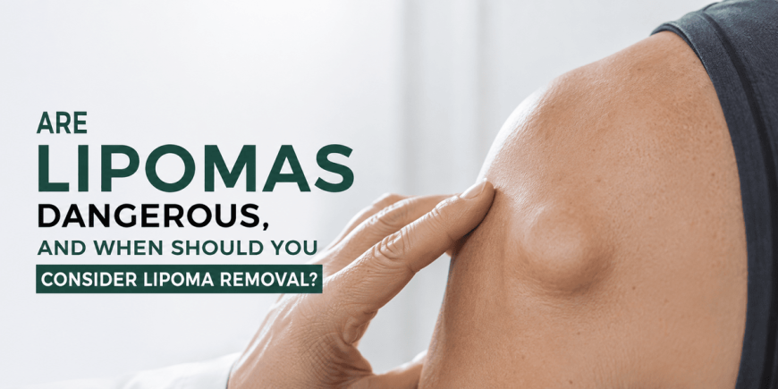
Most lipomas are harmless, but not every soft-tissue lump should automatically be assumed to be a lipoma. Knowing the typical signs and warning features can help you understand when assessment or lipoma removal may be appropriate. A soft, doughy lump under the skin on the shoulder, arm, or thigh often remains unchanged for months or years. Many people notice these growths during daily activities and wonder about possible risk. Lipomas are benign collections of mature fat cells that form in the subcutaneous tissue. They usually grow slowly, feel movable, and cause symptoms only when they press on nearby structures or become large. This article explains their nature, typical behaviour, and the situations that commonly prompt discussion of removal.
What Exactly Is a Lipoma?
A lipoma is a soft, benign growth made of mature fat cells (adipocytes) enclosed in a thin fibrous capsule. These lesions develop in the layer of tissue just beneath the skin.
They rank among the most frequent soft-tissue growths seen in adults. Most remain small, although some enlarge gradually over time. Lipomas are not infections or cysts. They differ from malignant tumours in both behaviour and cellular structure.
The exact reason a lipoma forms is not always clear. Genetic factors and, in some cases, minor local trauma appear to play a role. Recognising a lipoma as a localised overgrowth of ordinary fat tissue helps explain why many require no active treatment.
How Can You Recognise a Typical Lipoma?
Lipomas share several consistent physical features that help distinguish them from other soft-tissue lumps.
These characteristics support self-observation and guide decisions about seeking professional review.
- Texture and Consistency: Soft and doughy or rubbery. The lump often compresses slightly under gentle pressure and feels uniform.
- Mobility: Freely movable under the skin. It can usually be shifted from side to side without attachment to deeper structures.
- Growth Pattern: Slow change over months or years. Sudden or rapid enlargement is uncommon and warrants clinical assessment.
- Surface Appearance: The overlying skin typically looks normal, without redness, scaling, or a central pore.
- Size Variation: Lipomas vary considerably in size, and some can become much larger over time.
- Symptoms: Most cause no pain. Discomfort may occur if the growth presses on a nerve, limits movement, or sits in a high-friction area.
- Common Locations: Shoulders, upper back, arms, thighs, neck, and trunk—regions with more subcutaneous fat.
Clinical Insight
Dermatologists commonly observe that patients worry about firmness or recent growth. In practice, a soft, freely movable lump with normal overlying skin is characteristic of a lipoma. Any lesion that feels fixed, grows rapidly, or develops surface changes should be examined carefully because other soft-tissue tumours can sometimes resemble a lipoma.
Are Lipomas Dangerous or Linked to Cancer?
Most lipomas are benign and do not become cancerous. However, a new, rapidly growing, painful, firm, or fixed lump should be assessed by a qualified clinician because other types of soft-tissue tumours can resemble a lipoma.
True malignant change within a classic lipoma is very uncommon. Liposarcoma is a separate malignant fatty tumour that usually arises independently rather than from a pre-existing lipoma. Liposarcomas may grow faster, feel firmer, or cause pain, yet these features alone cannot reliably distinguish them from ordinary lipomas. Clinical examination, imaging, and, when needed, tissue analysis are required for accurate diagnosis.
Professional guidelines emphasise that typical soft, slow-growing, mobile lipomas do not need removal solely to prevent cancer. Concern arises mainly when the lesion deviates from the classic pattern. In those situations, further evaluation provides clarity and supports appropriate management.
What Factors Are Linked to Lipoma Development?
Several factors are associated with the appearance of lipomas, although a single cause is rarely identified in every case.
The strongest and most consistent associations include the following:
- Genetic Predisposition: Family history is frequently reported. Certain inherited patterns, such as familial multiple lipomatosis, lead to numerous lipomas.
- Age: Most lipomas become noticeable in middle adulthood, although they can appear at other ages.
- Occasional Local Trauma: Some individuals report a lipoma developing at the site of previous injury. This link is observational rather than proven in every case.
- Multiple Lesions: People who develop one lipoma sometimes develop others over time, consistent with an underlying predisposition.
Other associations occasionally appear in the medical literature, yet evidence varies in strength. Clinical focus remains on the features of the individual lump rather than on unproven systemic links.
When Might Lipoma Removal Be Considered?
Removal is usually an elective decision based on symptoms, size, location, diagnostic uncertainty, and personal preference.
Pain or discomfort from pressure on a nerve is a frequent reason for seeking treatment. Larger lipomas that restrict movement or interfere with clothing also prompt discussion. Cosmetic concern, especially when the lump sits in a visible area, motivates many people. Diagnostic uncertainty—when the lesion feels atypical—leads clinicians to recommend excision so that tissue can be examined in the laboratory. Rapid growth or a change in texture likewise justifies assessment and possible removal.
Lipoma removal treatment in London is therefore considered on an individual basis after careful evaluation of benefits, expected scarring, and recovery. Observation remains appropriate for small, stable, asymptomatic lipomas that show classic clinical features.
How Do Clinicians Confirm the Diagnosis?
Accurate diagnosis rests on clinical features and, when needed, additional investigations.
The process is usually straightforward for typical lipomas and more detailed when findings raise questions.
This structured approach ensures recommendations rest on reliable information rather than assumption.
What Treatment Options Are Available?
Once a decision for removal has been reached, several techniques may be considered. Suitability depends on the individual lesion, its location, and clinical assessment.
Surgical Excision
Surgical excision is the most widely used and reliable method. Under local anaesthetic, a small incision is made, the lipoma and its capsule are removed completely, and the wound is closed with fine sutures. This approach allows full laboratory examination of the tissue and carries a low risk of recurrence of the same lesion. It remains the primary technique for most lipomas that require removal.
Alternative Approaches
For selected larger or softer lipomas, liposuction-assisted techniques through small entry points may be discussed. These methods can reduce the length of the scar in some cases. However, the capsule may not always be fully removed, so residual tissue is possible. Suitability depends on the specific characteristics of the lipoma and the clinician’s judgement. Other minimally invasive variations exist but are used less frequently.
The clinician explains the advantages, limitations, and expected recovery of each option so that the patient can participate in the decision.
What Are the Benefits, Risks, and Limitations of Removal?
Every procedure balances potential advantages against possible drawbacks.
Benefits include relief of discomfort, improved contour, and diagnostic certainty when tissue is examined. Limitations include a small scar, temporary bruising or numbness, and the possibility that new lipomas may still form in other locations. Infection and fluid collection are uncommon when sterile technique and aftercare guidance are followed. Recurrence of the identical lipoma is uncommon after complete capsule removal.
Patients with bleeding tendencies or those taking anticoagulant medication require additional planning. Open discussion of these points supports realistic expectations. Observation remains a valid choice when the lipoma causes no symptoms and clinical features are typical.
Skin Health Snapshot
- Most lipomas remain stable and cause no symptoms for years.
What Happens Before, During, and After Removal?
Preparation, the procedure, and recovery follow a clear sequence for most people.
A consultation reviews medical history, examines the lipoma, and discusses options. On the treatment day the area is cleaned and local anaesthetic is given. The chosen technique is performed, typically lasting 20–60 minutes depending on size. A dressing is applied and written aftercare instructions are provided.
Mild soreness, bruising, or swelling is common for a few days and usually settles with simple measures. Sutures, when used, are removed according to the site, often within one to two weeks. Most people return to light activities within days and resume fuller exercise once the wound has healed. Sun protection on the scar supports a favourable cosmetic result. Follow-up allows review of healing and any laboratory findings.
At a Harley street dermatology clinic, clinicians routinely guide patients through these steps so that recovery proceeds smoothly and questions can be addressed promptly.
When Is Professional Assessment Particularly Important?
Certain features call for timely medical review rather than continued observation alone.
A new or changing lump should be assessed when its diagnosis is uncertain, particularly if it is growing, painful, firm, deep, or fixed. Surface changes, reduced mobility, or a lesion that feels markedly different from previous lipomas also justify examination. Individuals with a personal or family history of soft-tissue tumours may benefit from a lower threshold for review.
When uncertainty exists, clinical examination combined with imaging or excision provides clarity. Early assessment of atypical features supports accurate diagnosis while avoiding unnecessary procedures for classic, stable lipomas.
Illustrative Example
A person in their fifties noticed a soft, movable lump on the upper arm that had enlarged slowly over three years. Mild discomfort occurred during exercise. Clinical examination and ultrasound supported the diagnosis of a typical lipoma. After discussion of options, the lesion was excised under local anaesthetic. Laboratory analysis confirmed a benign lipoma with complete capsule removal. Recovery was uncomplicated, and normal activities resumed within a week. This example shows how characteristic clinical findings, supported by imaging when helpful, allow confident diagnosis and elective removal focused on symptom relief.
Comparing Lipomas with Other Common Soft-Tissue Lumps
|
Feature |
Typical Lipoma |
Epidermoid Cyst |
Other Soft-Tissue Tumours (including rare malignancies) |
Sebaceous Hyperplasia |
|
Consistency |
Soft, doughy, rubbery |
Firm to fluctuant |
Often firmer or more variable |
Soft, yellowish papules |
|
Mobility |
Freely movable |
Movable; may have central punctum |
May feel fixed or less mobile |
Surface-based |
|
Growth Rate |
Slow |
Slow; can inflame suddenly |
Variable; sometimes faster |
Very slow |
|
Pain |
Uncommon unless pressure on nerves |
Common if infected |
More variable |
None |
|
Overlying Skin |
Usually normal |
Possible central pore |
May show changes |
Yellowish, lobulated |
|
Cancer Risk |
Very low |
Negligible |
Present in malignant types |
Negligible |
|
Usual Management |
Observation or elective removal |
Observation or excision if symptomatic |
Prompt specialist assessment |
Observation or treatment if bothersome |
FAQs About Lipomas and Removal
Are lipomas a sign of a serious health condition?
In most cases, lipomas are isolated benign growths and do not indicate serious underlying disease. Multiple lipomas may prompt consideration of genetic predisposition, but the lumps themselves remain benign.
Can a lipoma turn into cancer?
Most lipomas do not become cancerous. Other soft-tissue tumours can occasionally resemble a lipoma, so any rapidly growing, painful, firm, or fixed lump should be assessed by a clinician.
Is lipoma removal always necessary?
No. Small, stable, asymptomatic lipomas with classic features can often be observed. Removal is considered when the lump causes symptoms, creates practical difficulty, raises diagnostic questions, or causes significant cosmetic concern.
What does recovery after removal involve?
Most procedures use local anaesthetic. Mild bruising and soreness usually settle within days to a week. Sutures are removed according to the site, and normal activities resume progressively as the wound heals.
Will new lipomas appear after one is removed?
Removal addresses the existing lesion. New lipomas can still develop in other locations. Complete capsule excision makes recurrence of the same lipoma uncommon.
To Bring It All Together
Lipomas are common benign fatty growths that form under the skin and usually enlarge slowly. Most remain harmless and require no treatment. Recognition of their soft, movable nature allows many people to monitor them with confidence. Removal becomes a reasonable consideration when the lipoma causes discomfort, interferes with movement or clothing, raises diagnostic uncertainty, or creates significant cosmetic concern. Clinical examination, sometimes supported by ultrasound, guides the decision and the choice of technique. Surgical excision under local anaesthetic remains the most reliable method when intervention is chosen, providing both symptom relief and tissue confirmation. Ongoing awareness of any new or changing lumps supports long-term soft-tissue health.
Medical Review
This article is intended for general educational purposes. Medical information should be reviewed periodically in light of current clinical guidelines.